Relying on 20/20 vision as a sign of perfect eye health is a dangerous oversimplification; it measures only one aspect of a complex biological system.
- A comprehensive exam is a proactive data-gathering process that maps your unique “biological blueprint,” detecting silent shifts in your ocular and systemic health.
- Conditions like glaucoma, diabetic retinopathy, and even hypertension often show their first signs in the eye, long before symptoms appear.
Recommendation: Treat your routine eye exam not as a pass/fail vision test, but as an essential part of your preventive health strategy, just like a physical or dental check-up.
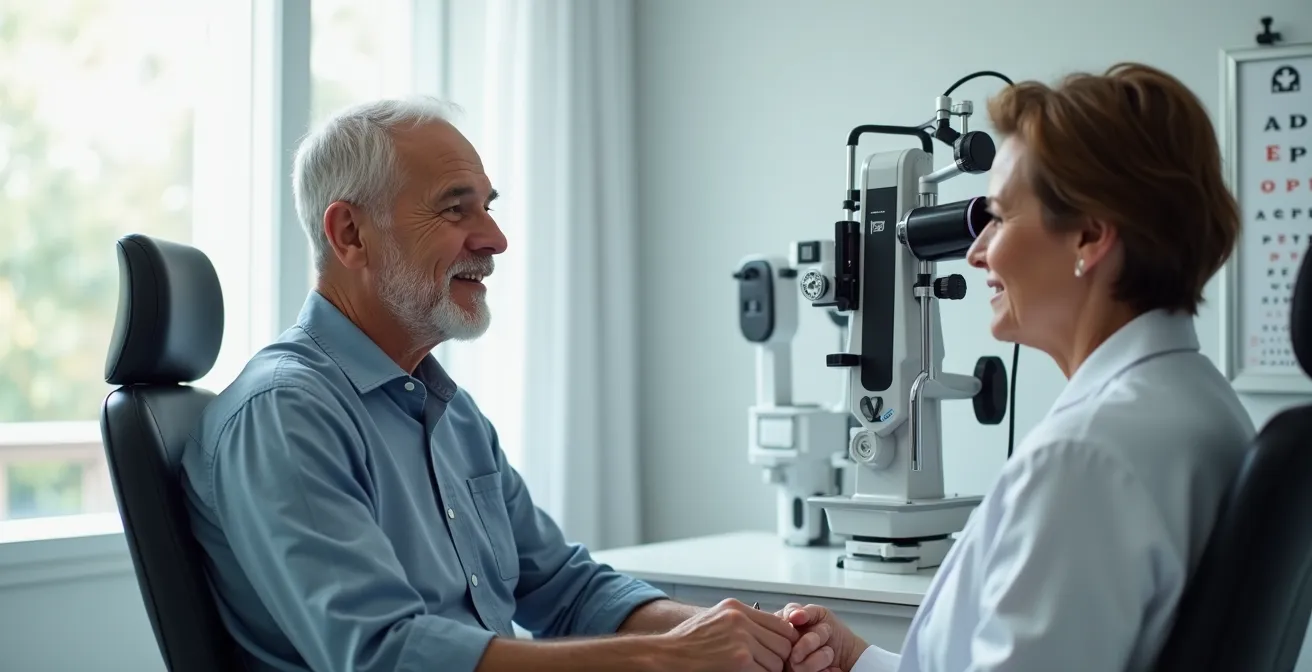
If you can read the bottom line of an eye chart without trouble, you might feel invincible. For many adults, especially those in their 30s with no corrective lenses, the idea of a routine eye exam seems redundant. The common thinking is, “If I can see perfectly, my eyes must be perfectly healthy.” This logic is simple, understandable, and dangerously flawed. Perfect vision is not a proxy for perfect health, and the belief that it is leaves millions of people vulnerable to preventable, vision-threatening conditions.
The standard advice to “get your eyes checked” often gets lost in a sea of public health messaging. We’re told it detects “silent” diseases, but for someone who feels healthy, this warning can seem abstract and distant. But what if we reframe the question? What if an eye exam isn’t just a test you pass or fail? The truth is, a comprehensive eye exam is a sophisticated, data-gathering process. It’s less about a score and more about mapping the unique biological blueprint of your visual system, revealing subtle changes and future risks that a simple acuity test could never identify.
This article will deconstruct the myth of 20/20 vision as a clean bill of health. We will explore the specific, often invisible, signs that only a thorough examination can detect—from the structural integrity of your retina to the first clues of systemic diseases like diabetes and hypertension. We will analyze why even perfect daytime vision can betray you at night, what modern technology reveals about your eyes, and why an in-office exam is an irreplaceable diagnostic tool for your overall well-being.
For those who prefer a visual format, the following video offers a brief history of the standard eye chart and the origins of the 20/20 measurement, providing a perfect complement to the detailed information in this guide.
This guide provides a comprehensive overview of why a routine eye exam is a cornerstone of preventive health, even for those with seemingly perfect sight. Below, you will find a detailed breakdown of the key topics we will cover.
Summary: Why a Routine Eye Exam Is Non-Negotiable, Even with 20/20 Vision
- Why 20/20 Vision Is Not a Clean Bill of Health?
- Why Your 20/20 Vision Blurs While Driving at Night?
- Why Skipping Dilation Might Cost You Your Peripheral Vision?
- How Retinal Imaging Replaces the Ophthalmoscope in Modern Exams?
- The Danger of Hiding Your Hypertension Medication From Your Eye Doctor
- When to Schedule the First Exam: Before Reading or Before Walking?
- Online Vision Test or In-Office Exam: Which Is Safe for Prescription Renewals?
- When to Schedule Your Eye Exam: Morning vs. Afternoon Accuracy
Why 20/20 Vision Is Not a Clean Bill of Health?
The term “20/20 vision” is one of the most recognized benchmarks in healthcare, yet it’s widely misunderstood. It simply means you can see at 20 feet what a person with normal vision can see at 20 feet. It is a measurement of visual acuity, or sharpness, at a specific distance. However, it is not a comprehensive evaluation of your eye health. In fact, according to the American Academy of Ophthalmology, only about 35% of all adults have 20/20 vision without any form of correction. The other 65% rely on glasses, contacts, or surgery to reach this standard.
The critical mistake is equating this single data point with overall health. As Dr. Mark Wilkinson of the University of Iowa Carver College of Medicine explains, the 20/20 measurement is merely a point of reference:
The 20/20 mark is ‘simply a point of reference.’ It doesn’t speak to quality of vision, which is one of the things that people get confused about — quality versus quantity. The 20/20 measurement gauges the quantity of your vision (the ability to recognize letters on an eye chart), while a contrast sensitivity test determines the quality.
– Dr. Mark Wilkinson, University of Iowa Carver College of Medicine
Your visual system is far more complex than just acuity. It includes peripheral vision, eye coordination, depth perception, color vision, and focusing ability. More importantly, many of the most serious eye diseases, such as glaucoma, diabetic retinopathy, and macular degeneration, can develop for years without affecting your central visual acuity. A person can have 20/20 vision and simultaneously be losing their peripheral sight to glaucoma, a condition that causes irreversible damage if not caught early.
The following table illustrates several sight-threatening conditions that often have no impact on 20/20 vision in their early stages, highlighting why a comprehensive exam is so vital.
| Eye Condition | Early Signs Often Missed | Potential Consequences |
|---|---|---|
| Glaucoma | No symptoms initially; peripheral vision loss | Permanent optic nerve damage |
| Macular Degeneration | Subtle central vision changes | Loss of central vision |
| Diabetic Retinopathy | No early symptoms | Vision loss from blood vessel damage |
| Retinal Detachment | Small tears in periphery | Permanent vision loss if untreated |
Why Your 20/20 Vision Blurs While Driving at Night?
One of the most common complaints from people with otherwise perfect daytime vision is a noticeable decline in visual quality at night. Headlights from oncoming cars create starbursts or halos, road signs seem less distinct, and judging distances becomes a challenge. This isn’t just an inconvenience; it’s a significant safety issue. An analysis by the AAA Foundation for Traffic Safety reveals that while only 25% of automotive travel occurs at night, this period accounts for a staggering 50% of all driver fatalities.

So why does this happen? The answer lies in how your visual system adapts to low-light conditions. When it gets dark, your pupils dilate to let in more light. This process can expose subtle optical imperfections in your eye’s lens and cornea, known as higher-order aberrations, which go unnoticed in bright light when your pupils are smaller. These imperfections scatter light, causing the classic symptoms of glare and halos. Furthermore, conditions like early-stage cataracts or uncorrected astigmatism are much more pronounced in the dark.
Another often-overlooked cause is Binocular Vision Dysfunction (BVD). This condition, where the eyes are slightly misaligned, affects an estimated 56% of American adults. During the day, your brain can easily compensate for the misalignment. But at night, the visual system is already under stress. The extra effort required to fuse the two slightly different images from your eyes can lead to symptoms like double vision, shadowed text on signs, and eye strain. An in-office exam is the only way to test for and diagnose BVD, as it requires specific tests of eye alignment and coordination that go far beyond a simple acuity chart.
Why Skipping Dilation Might Cost You Your Peripheral Vision?
For many, the most dreaded part of an eye exam is pupillary dilation. The eye drops, the light sensitivity, and the blurry up-close vision for a few hours can feel like a major hassle. It’s tempting to ask to skip it, especially if you feel your vision is fine. However, refusing dilation is like asking a mechanic to inspect your car’s engine without opening the hood. Your pupil is a window to the back of your eye, and dilation simply opens that window wide, allowing your doctor to get an unobstructed, three-dimensional view of your optic nerve, retina, and blood vessels.
This view is absolutely critical for detecting serious conditions in their earliest stages. Glaucoma, often called the “silent thief of sight,” damages the optic nerve and slowly erodes your peripheral vision, often without any symptoms until significant, irreversible vision loss has occurred. A dilated exam allows the optometrist to examine the optic nerve’s shape, color, and health to spot the subtle cupping and notching characteristic of early glaucomatous damage.
Similarly, a dilated view of the peripheral retina is essential for finding retinal tears or detachments, which can occur from injury or simply age. These often start as tiny defects on the outer edges of the retina—areas completely invisible through an undilated pupil. A study from the New England College of Optometry found that combining modern imaging with a dilated examination increased overall retinal lesion detection by 30% compared to older methods. Skipping dilation means these early warnings can be missed entirely, with potentially devastating consequences for your sight.
How Retinal Imaging Replaces the Ophthalmoscope in Modern Exams?
The traditional tool for looking into the eye is the ophthalmoscope, a handheld device that projects a small beam of light. While effective, it provides a very narrow field of view, much like looking at a large mural through a keyhole. Modern optometry has revolutionized this process with ultra-widefield (UWF) retinal imaging. This technology uses a digital scanning laser to capture a panoramic image of the retina in a single, high-resolution shot, often in less than a second.
A UWF image can capture up to 200 degrees of the retina, compared to the 15-60 degrees visible with traditional methods. This expansive view allows doctors to see the far periphery of the retina, where signs of problems like diabetic retinopathy, retinal tears, and vascular occlusions often first appear. The high-resolution digital image can be magnified to inspect tiny details of the optic nerve and macula. Most importantly, these images create a permanent digital record of your eye’s internal structures. This establishes a baseline “biological blueprint” of your retina, allowing your doctor to track subtle, year-over-year changes with an incredible degree of precision.
The power of this data is now being amplified by artificial intelligence. As the Poudre Valley Eyecare Research Team notes, AI is reaching new heights in diagnostic accuracy:
In April 2024, AEYE-DS became the first fully autonomous AI algorithm to receive FDA clearance for diagnosing referable diabetic retinopathy from retinal images taken by handheld cameras. This breakthrough technology achieved sensitivity rates of 92-93% and specificity rates of 89-94% in large-scale clinical trials.
– Poudre Valley Eyecare Research Team, Mass Eye and Ear Predictive Health Research Study
This fusion of imaging and AI embodies the shift from reactive problem-solving to proactive health mapping. It is a powerful tool in your doctor’s arsenal, transforming the eye exam into a high-tech data collection session.
The Danger of Hiding Your Hypertension Medication From Your Eye Doctor
The eye is the only place in the body where blood vessels can be observed directly, non-invasively. This unique window makes an eye exam a powerful tool for detecting signs of systemic health conditions, sometimes even before they are diagnosed by a primary care physician. Conditions like diabetes, high cholesterol, autoimmune disorders, and even certain types of brain tumors can leave tell-tale signs on the retina or optic nerve.
One of the most common and critical connections is with hypertension, or high blood pressure. Chronic high blood pressure can damage the delicate blood vessels in the retina, causing them to leak, narrow, or harden. This condition, known as hypertensive retinopathy, can lead to blurred vision or even permanent vision loss if left unmanaged. During a dilated exam, your optometrist can see these changes directly and assess their severity. This information is not only vital for your eye health but also serves as a crucial indicator of how well your blood pressure is being controlled throughout your entire body.
This is why it is extremely dangerous to withhold information about your medications from your eye doctor. Some patients might be embarrassed or think it’s irrelevant, but your doctor needs the full picture. If they see changes in your retinal blood vessels, knowing you are on hypertension medication helps them determine if the current treatment is effective or if a discussion with your primary doctor is needed. As one patient was told regarding vision changes, “If you have noticed that your vision is blurry, cloudy, dim, or if you notice ‘halo effects,’ you’ll want to address it right away.” These symptoms could be linked to blood pressure fluctuations, and your eye doctor is on the front lines of spotting the connection.
When to Schedule the First Exam: Before Reading or Before Walking?
The concept of preventive eye care should begin long before a child can read an eye chart. A significant amount of a child’s visual development occurs in the first few years of life, and early detection of problems is critical to ensure proper development and prevent lifelong vision issues. Conditions like strabismus (crossed eyes) and amblyopia (lazy eye) are most effectively treated when identified at a very young age. If left untreated, the brain may learn to ignore the input from the weaker eye, leading to permanent vision impairment.
Because of this, experts recommend a child’s first comprehensive eye exam occur much earlier than most parents realize. It’s not about testing their ability to read, but about assessing the fundamental health and alignment of their eyes. According to the American Optometric Association, the timeline for eye exams is a lifelong commitment:
The American Optometric Association recommends that adults aged 18 to 60 should have an eye exam at least every two years, and annually for those over 60. Children should have their first eye exam at 6 months, then at age 3, and before starting school, followed by regular exams as their eye doctor recommends.
– American Optometric Association, Comprehensive Eye and Vision Examination Guidelines
The exam at six months is particularly crucial. During this visit, an optometrist will check for proper eye movement, alignment, and signs of congenital cataracts or other structural abnormalities. This early screening establishes a baseline and ensures the child’s visual system is on the right developmental track. Thinking of eye care as a process that begins in infancy, rather than when a problem is noticed, reframes the exam as a fundamental pillar of pediatric healthcare, just as important as vaccinations and well-child visits.
Online Vision Test or In-Office Exam: Which Is Safe for Prescription Renewals?
In our digital-first world, the convenience of online vision tests for renewing a glasses or contact lens prescription is undeniably appealing. These services allow you to use your smartphone or computer to read an eye chart, and a remote doctor approves the prescription. While this may seem like an efficient alternative, it is not a substitute for a comprehensive, in-office eye exam. An online test only measures one thing: visual acuity. It completely bypasses the most critical component of an eye exam—the health assessment.
An in-office exam is a medical assessment, not just a refraction. Optometrists are trained to detect a vast array of health issues that have no early symptoms. They are often the first healthcare providers to spot signs of serious systemic diseases. For example, a 2018 report found that optometrists detected over 301,000 cases of diabetes in patients who had not yet been diagnosed by their primary physician. This is possible because diabetes causes characteristic changes to the blood vessels in the retina, which are only visible during a comprehensive exam.
By opting for an online test, you are choosing convenience over your health and safety. You are forgoing the vital checks that can catch sight-threatening and even life-threatening conditions early. The difference in what is evaluated is stark, and understanding this difference is key to making an informed decision about your health care.
Action Plan: Key Checks Performed in a Comprehensive Exam
- Visual acuity test to measure clarity at different distances plus refraction testing for exact prescription.
- Eye movement and coordination assessment for reading and computer tasks requiring precise vision.
- Intraocular pressure measurement using tonometry for early glaucoma detection.
- Dilated pupil examination for checking the retina, optic nerve, and blood vessels for conditions like diabetic retinopathy and macular degeneration.
- Slit lamp examination of the cornea, iris, and lens for detecting cataracts and corneal damage.
Key Takeaways
- 20/20 vision only measures sharpness; it says nothing about your peripheral vision, eye pressure, or retinal health.
- A comprehensive exam is a data-driven process that can reveal the first signs of glaucoma, diabetes, and hypertension years before other symptoms appear.
- Modern tools like ultra-widefield retinal imaging create a permanent digital record of your eye, allowing for precise tracking of subtle health changes over time.
When to Schedule Your Eye Exam: Morning vs. Afternoon Accuracy
Once you’ve committed to a comprehensive eye exam, a final practical question arises: does the time of day matter? The answer is yes, it can. Your body, including your eyes, is subject to daily rhythms that can influence the results of your exam. For the most accurate refraction—the part of the exam that determines your precise prescription for glasses or contacts—it is generally best to schedule your appointment for a time when your eyes are not fatigued.
For many people, this means a morning appointment is ideal. After a full night’s rest, your eyes are fresh. The muscles that control focusing have not been strained by hours of staring at a computer screen or reading documents. As the day wears on, eye strain can set in, potentially leading to a slight over-correction in your prescription, as your tired focusing system works harder than it should. This is sometimes referred to as “pseudomyopia,” where eye fatigue mimics nearsightedness.
Other factors can also skew results. If you suffer from allergies, your eyes may be more irritated and watery in the morning or evening, which could interfere with the exam. Similarly, performing strenuous exercise right before your appointment can temporarily affect your eye pressure readings. The best approach is to communicate with your doctor’s office. Mention your daily work habits and any known issues like eye strain or allergies. They can help you choose an appointment time when your eyes are most likely to be in their natural, rested state, ensuring the data collected during your exam is as accurate as possible.
Frequently Asked Questions About How Often Should You Get an Eye Exam if You Have 20/20 Vision?
Does the time of day affect eye exam results?
Yes, your eye exam results can change depending on the time of day. Results may be skewed if you are tired, have just exercised, or are experiencing allergy symptoms, as these factors can cause temporary changes in your vision and eye pressure.
What activities should be avoided after dilated eye exams?
After your pupils are dilated, your eyes will be very sensitive to light and your near vision will be blurry. You should avoid driving, spending time in bright sun without sunglasses, and trying to focus on digital screens or books for several hours until the effects wear off.
How long do eyes stay dilated after an exam?
First, you have to wait 20-30 minutes for the dilating drops to take full effect before the doctor can perform the examination. The effects of dilation, including light sensitivity and blurry near vision, typically last for 4 to 6 hours, though this can vary from person to person.