Optics and vision
Your eyes are among the most sophisticated optical instruments in nature, yet many people navigate vision care without fully understanding how their eyes work or what their test results actually mean. From the seemingly simple Snellen chart hanging in your eye doctor’s office to the complex prescription slip you receive, the science of optics and vision encompasses a rich landscape of measurements, technologies, and biological processes that directly impact your daily visual experience.
Whether you’re considering your first pair of glasses, puzzled by fluctuating prescription values, or simply curious about how contact lenses differ from spectacles, grasping the fundamentals of vision science empowers you to make informed decisions about your eye health. This comprehensive resource breaks down the essential concepts—from visual acuity and prescription parameters to corneal breathability and advanced imaging techniques—in clear, practical terms that demystify the numbers and terminology you encounter at every eye examination.
Understanding Visual Acuity: What Your Eyes Can Really See
Visual acuity represents your eye’s ability to discern fine details at a specific distance, but this single measurement tells only part of the story about your functional vision. The familiar 20/20 standard has become synonymous with “perfect vision” in popular culture, yet this benchmark actually describes average normal vision rather than the theoretical limit of human visual capability.
The Science Behind the Numbers
When an optometrist references 20/20 vision, they’re describing a ratio: what you can see clearly at 20 feet is what a person with statistically normal vision should see at that same distance. Someone with 20/40 vision must stand at 20 feet to see what normal vision sees at 40 feet. This measurement system, based on the Snellen chart with its progressively smaller letters, has limitations—it tests only high-contrast static targets under optimal lighting conditions.
In reality, your visual performance varies dramatically based on environmental factors. Low light conditions reduce acuity because your eye’s photoreceptors switch from cones (which provide sharp color vision) to rods (which offer less resolution but greater light sensitivity). This explains why reading fine print becomes challenging at dusk even if you have excellent daytime acuity.
Static Versus Dynamic Vision
Standard acuity tests measure static visual acuity—your ability to see stationary objects—but daily life demands dynamic visual acuity as well. Tracking moving objects, reading while walking, or identifying road signs from a moving vehicle all require different visual processing. Athletes and drivers particularly benefit from understanding this distinction, as static test results don’t necessarily predict performance in motion-dependent scenarios.
When and Why to Schedule Re-evaluations
Visual acuity isn’t permanently fixed. Children’s vision develops until their late teens, while adults experience gradual changes due to aging, health conditions, or environmental factors. Scheduling regular evaluations—typically annually for adults and more frequently for children or those with existing conditions—allows for timely prescription adjustments before vision changes significantly impact daily activities. Skipping the dilation portion of these exams, though temporarily more convenient, can cause practitioners to miss subtle refractive errors or early signs of eye disease.
Decoding Your Prescription: Numbers That Define Your Vision
An eyeglass prescription resembles a mathematical formula filled with positive and negative numbers, abbreviated terms, and measurements that can seem cryptic without context. Yet each value precisely describes how light must be bent to focus correctly on your retina, compensating for your eye’s unique optical characteristics.
Why Prescription Values Fluctuate
You might notice slightly different measurements between examinations or even at different times of day. This variation occurs because multiple factors influence refraction testing. Your eye’s focusing muscles experience fatigue throughout the day, particularly after extended near work. Hydration levels affect the cornea’s curvature slightly, while blood sugar variations can temporarily alter the lens shape. These fluctuations typically remain within 0.25 to 0.50 diopters—a range most people don’t notice functionally.
Verifying Your Lenses Match the Prescription
Receiving new glasses should improve your vision, but manufacturing errors occasionally occur. If your new eyewear feels uncomfortable or vision seems wrong, opticians can verify the lenses using a lensometer, which measures the actual optical power. This quick, non-invasive test confirms whether the laboratory accurately produced lenses matching your prescription specifications.
Material Choices: High Index Versus Polycarbonate
Lens materials represent a balance between optical clarity, weight, thickness, and impact resistance. Polycarbonate lenses offer exceptional durability and built-in UV protection, making them ideal for children, athletes, and safety glasses. High-index materials bend light more efficiently, allowing thinner, lighter lenses for strong prescriptions, though at a higher cost. Your specific prescription strength, lifestyle needs, and budget should guide this choice rather than a one-size-fits-all approach.
The Hidden Costs of Expired Prescriptions
Using an outdated prescription might seem harmless if your vision “feels fine,” but expired prescriptions carry risks beyond suboptimal clarity. Prescriptions expire because eye health changes over time—sometimes subtly. Wearing incorrect correction can cause eye strain, headaches, and visual fatigue. More critically, regular examinations detect conditions like glaucoma, cataracts, or retinal problems in early stages when treatment proves most effective.
Diopters Explained: The Universal Language of Optical Power
The diopter serves as the fundamental unit of measurement for optical power, quantifying how strongly a lens bends light. Understanding diopters unlocks the logic behind prescription numbers and clarifies why certain corrections work differently for various vision problems.
Positive Versus Negative Diopters
A lens measured at +2.00 diopters converges light rays to a focal point, correcting farsightedness (hyperopia) where the eye focuses images behind the retina. Conversely, a -2.00 diopter lens diverges light, correcting nearsightedness (myopia) where images focus in front of the retina. This positive/negative distinction fundamentally describes whether the lens adds or subtracts focusing power to compensate for your eye’s natural refractive error.
Converting Between Contact Lenses and Glasses
Contact lens prescriptions differ numerically from eyeglass prescriptions for the same person because contacts sit directly on the cornea while glasses rest roughly 12 millimeters from the eye. This distance difference, called vertex distance, becomes significant for prescriptions stronger than ±4.00 diopters. A simple formula adjusts for this geometric relationship, which is why you cannot simply transfer numbers from your glasses prescription to order contacts—each requires its own fitting and measurement.
The Limits of Surgical Correction
Refractive surgeries like LASIK reshape the cornea to alter its optical power, but anatomical constraints limit how much correction is safely possible. The cornea must retain sufficient thickness for structural integrity, typically restricting correction to approximately -12.00 diopters of myopia or +6.00 diopters of hyperopia. Extreme prescriptions may require alternative approaches like phakic intraocular lenses or refractive lens exchange.
Why Diopters Don’t Directly Equal Visual Acuity
A common misconception equates diopter measurements with acuity scores, but these represent entirely different concepts. Diopters measure optical power needed for correction; visual acuity measures the result of that correction. Someone with -3.00 diopters of myopia might achieve 20/20 acuity with proper correction, while another person with the same prescription might reach only 20/30 due to other factors like astigmatism, amblyopia, or retinal conditions that glasses cannot correct.
Corneal Oxygen: Why Your Eyes Need to Breathe
Unlike most body tissues, your cornea lacks blood vessels to deliver oxygen and nutrients. Instead, it relies on atmospheric oxygen dissolving through the tear film, making breathability a critical consideration for anyone wearing contact lenses that create a barrier between air and the corneal surface.
The cornea’s oxygen requirements remain constant whether you’re awake or asleep. When oxygen supply drops below necessary levels, a condition called hypoxia develops, triggering a cascade of responses. Early signs include excessive tearing, redness, and slight blurring that improves after removing lenses. Chronic oxygen deprivation can stimulate abnormal blood vessel growth (neovascularization) into the normally clear cornea, permanently affecting vision and reducing future contact lens options.
Modern lens materials address this challenge through varying approaches. Traditional hydrogel lenses allow oxygen to dissolve in their water content—higher water percentage generally means better oxygen transmission but may reduce durability. Newer silicone hydrogel materials incorporate silicone polymers that permit oxygen to pass directly through the lens matrix, achieving dramatically higher oxygen permeability even with lower water content. This technological advancement has made extended wear safer, though no lens transmits oxygen as effectively as an unobstructed cornea.
Timing your lens wear appropriately protects corneal health long-term. Removing lenses before napping—even brief naps—prevents the compounded oxygen restriction that occurs when closed eyelids combine with lens barrier effects. Following manufacturer recommendations for daily wear limits, typically ranging from 12 to 16 hours depending on lens type, maintains the delicate balance between convenience and corneal wellness.
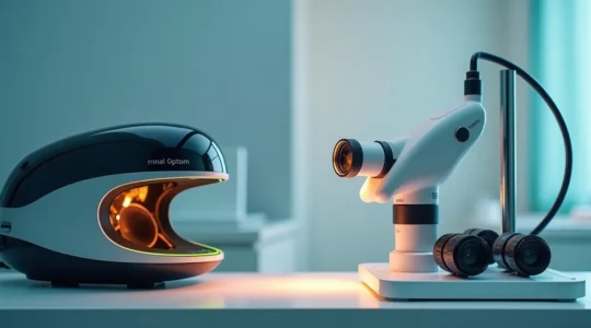
Retinal Imaging: Seeing Beyond the Surface
While traditional eye examinations assess refractive error and external eye health, advanced imaging technologies enable practitioners to visualize and analyze internal structures with unprecedented detail, detecting subtle changes long before they affect your conscious vision.
Optical Coherence Tomography (OCT) functions somewhat like ultrasound but uses light waves instead of sound, creating cross-sectional images of retinal layers with resolution approaching cellular level. Interpreting an OCT scan reveals the thickness and integrity of structures like the macula—responsible for central detailed vision—and can identify early macular degeneration, diabetic retinopathy, or glaucomatous damage affecting the optic nerve. These scans provide objective measurements that track disease progression or treatment effectiveness over time.
Fundus photography captures wide-angle color images of the retina’s surface, documenting the appearance of blood vessels, the optic disc, and the macula. While a live examination through dilated pupils allows dynamic viewing and depth perception, fundus photos create permanent records for comparison at future visits. The peripheral retina—areas outside the central viewing field—matters significantly because many serious conditions like retinal tears or peripheral tumors develop in these outer regions that are difficult to examine without dilation.
The bright flash accompanying retinal photography often concerns patients, spawning myths about potential damage, but these flashes operate well within safe exposure limits. The intensity feels dramatic because your pupils are dilated and light-sensitive, but modern imaging systems carefully regulate flash duration and energy to eliminate risk while capturing necessary detail.
Understanding warning signs that warrant urgent imaging helps preserve vision. New flashers (brief streaks of light in peripheral vision) or floaters (spots or strands drifting through your visual field) can indicate vitreous detachment or retinal tears. While many floaters represent benign age-related changes, the sudden onset of multiple floaters, especially accompanied by flashes or a shadow in peripheral vision, requires prompt evaluation—ideally within 24 hours—since retinal tears can progress to detachment if untreated.
This foundation in optics and vision science transforms confusing numbers and unfamiliar terminology into comprehensible information that supports better eye care decisions. Whether evaluating prescription changes, selecting contact lens materials, or understanding recommended testing, you now possess the conceptual framework to engage meaningfully with your vision care provider and advocate effectively for your ocular health.
Optomap or Dilation: Which Retinal Exam Is Right for You?
The decision between Optomap and dilation is not about comfort versus discomfort; it’s a strategic choice about the depth and breadth of diagnostic information you want to capture about your eye health. Wide-field imaging like Optomap excels at creating a…
Read more
Low Dk/t vs. High Dk/t: How Oxygen Permeability Affects Your Red Eyes?
The chronic redness and fatigue you feel from your contact lenses are not just discomfort; they are symptoms of an accumulating “physiological debt” in your cornea caused by oxygen starvation. Low oxygen permeability (low Dk/t) forces your cornea into an…
Read more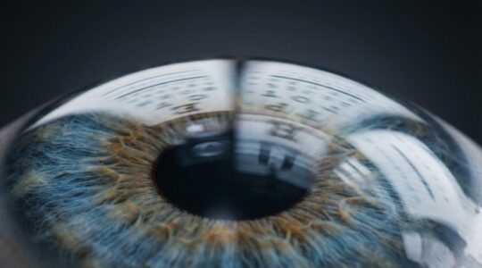
What Do -4.00 Diopters Actually Mean for Your Focal Distance?
Contrary to common belief, a -4.00 diopter prescription is not just a score for “bad vision”; it’s a precise physical measurement. It means your eye focuses light 25 centimeters in front of where it should, and the corrective lens required…
Read more
How to Confidently Read Your Eyeglass Prescription and Order Online
In summary: Your prescription is a precise map of your eye’s unique needs, not just a list of numbers. Understanding the “why” behind SPH, CYL, and AXIS is key. Factors like the time of day can affect your exam results,…
Read more
Understanding 20/20 Vision: Why Perfect Acuity Doesn’t Guarantee Eye Health
A 20/20 vision score is not a guarantee of perfect vision or complete eye health. It only measures your ability to see static, high-contrast objects from a distance and fails to test crucial functions like night vision, motion tracking, or…
Read more