A 20/20 vision score is not a guarantee of perfect vision or complete eye health.
- It only measures your ability to see static, high-contrast objects from a distance and fails to test crucial functions like night vision, motion tracking, or peripheral awareness.
- Serious, sight-threatening diseases like glaucoma can develop and destroy a significant portion of your vision without ever affecting your 20/20 score until the damage is irreversible.
Recommendation: If you experience visual discomfort, strain, or headaches despite a “perfect” test result, trust your symptoms. The next step is to request a comprehensive eye health exam that investigates beyond the standard chart.
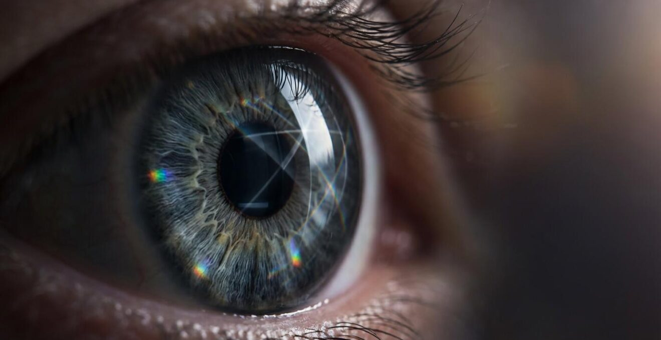
You’ve just left the optometrist’s office, certificate in hand, confirming you have 20/20 vision. Yet, the relief is fleeting. Why do you still struggle to see road signs clearly at night? Why does hours of screen time leave your eyes feeling strained and tired? And why do you sometimes feel a vague sense of visual unease that you can’t quite describe? This experience, a frustrating gap between a “perfect” test score and your real-world vision, is what I call the visual disconnect. It’s a common and valid concern that patients bring to me daily.
Most discussions around vision focus on the familiar Snellen eye chart and the goal of achieving 20/20 acuity. We are conditioned to believe this number is the ultimate benchmark of ocular health. However, this is a dangerous oversimplification. Your visual system is far more complex than its ability to identify stationary black letters on a white background. It encompasses depth perception, color recognition, peripheral awareness, and the ability to adapt to changing light conditions.
But what if the very tool we use to measure “perfect” vision is also creating an illusion of safety? The fundamental flaw in relying solely on a 20/20 score is that it measures only one specific function—static visual acuity—while ignoring the dynamic, functional needs of your eyes. This article will deconstruct that illusion. We will explore precisely why your perfect score can coexist with tangible discomfort, what silent deficits standard tests miss, and what a truly comprehensive view of your eye health should entail.
This guide is structured to walk you through the limitations of the standard eye test and empower you with the knowledge to advocate for your own eye health. We will cover the specific phenomena behind common visual complaints, explain how to interpret your results beyond the bottom line, and clarify why diagnostic tests that seem inconvenient are, in fact, essential shields against irreversible vision loss.
Summary: Deconstructing the Myth of Perfect 20/20 Vision
- Why Your 20/20 Vision Blurs While Driving at Night?
- How to Read Your Eye Chart Results Beyond the Bottom Line?
- Static vs. Dynamic Acuity: Which Matters More for Tennis Players?
- The Silent Retinal Issue That Standard Acuity Tests Miss in 1 out of 5 Patients
- When to Retest Vision: 3 Signals That Your Prescription Changed Early
- The Mistake of Assuming -1.00 Diopter Equals 20/40 Vision
- Why You Can Lose 40% of Your Vision Without Noticing It?
- Is the “Air Puff” Test Really Necessary During Your Annual Refractive Exam?
Why Your 20/20 Vision Blurs While Driving at Night?
One of the most common complaints from patients with excellent daytime acuity is a noticeable drop in visual quality at night, particularly while driving. This isn’t your imagination; it’s a physiological reality that the standard 20/20 test is simply not designed to detect. The Snellen chart exam is conducted in photopic conditions—bright, office-level lighting. Driving at dusk or at night, however, places your eyes in mesopic conditions, a state of intermediate light where both your cones (for color and detail) and rods (for low-light vision) are active.
In this state, a different visual function becomes paramount: contrast sensitivity. This is your ability to distinguish an object from its background, such as seeing a pedestrian in dark clothing against a shadowy street. An eye chart tests your ability to see high-contrast black letters on a white background. Night driving involves low-contrast scenarios where glare from oncoming headlights can further reduce your visual performance. Research confirms this diagnostic gap. As one study in the Journal of Optometry found, mesopic visual acuity and disability glare index are most strongly associated with night driving difficulties, not standard acuity.
This means you can have flawless 20/20 vision and still have poor contrast sensitivity, leading to that feeling of blurriness and insecurity on the road. A case study on driver performance reinforces this, showing that contrast sensitivity measured under mesopic conditions was a far better predictor of nighttime hazard detection than standard acuity tests. If you experience this, it is a clear signal that your functional vision needs assessment beyond the eye chart.
How to Read Your Eye Chart Results Beyond the Bottom Line?
The final line you read on the eye chart, whether it’s 20/20 or 20/40, is only the headline of your visual story. Your full prescription and exam notes contain a wealth of information that offers a much more nuanced picture of your eye health. Understanding these notations can empower you to have more informed conversations with your eye care professional and demystify the numbers and letters on your report.
The most common terms you’ll encounter are OD, OS, and OU. These are Latin abbreviations: OD (Oculus Dexter) refers to your right eye, OS (Oculus Sinister) to your left eye, and OU (Oculus Uterque) to both eyes working together. Your acuity may differ between each eye, and the OU measurement indicates how well your brain combines the two images. For instance, you could have 20/25 in your right eye and 20/20 in your left, with a combined vision of 20/20.
Another critical notation is the PH (Pinhole) test result. During this test, you look at the eye chart through a small pinhole. If your vision improves significantly, it strongly indicates that your vision problem is a simple refractive error (like nearsightedness or astigmatism) that can be corrected with glasses. If your vision does *not* improve with the pinhole, it is a red flag suggesting that the issue may be rooted in an underlying eye health problem, such as a cataract or a retinal issue, which requires further investigation.
Finally, it’s important to understand what the numbers themselves mean. A score of 20/40 means that you must be at 20 feet to see what a person with “normal” vision can see at 40 feet. Conversely, vision better than average, such as 20/15, means you can see clearly at 20 feet what a normal eye can only see at 15 feet. Here are the key notations to look for:
- OD (Oculus Dexter): Your right eye measurements
- OS (Oculus Sinister): Your left eye measurements
- OU (Oculus Uterque): Both eyes together
- PH (Pinhole) test result: If vision improves with pinhole, indicates refractive error; if not, suggests an eye health issue
- 20/40 notation: You see at 20 feet what normal vision sees at 40 feet
Static vs. Dynamic Acuity: Which Matters More for Tennis Players?
The standard eye chart measures static visual acuity—your ability to see stationary objects with clarity. This is useful for tasks like reading a book or a sign. However, for any activity involving motion, a completely different skill comes into play: dynamic visual acuity (DVA). This is your ability to see, track, and identify objects that are moving. For an athlete like a tennis player, DVA is arguably far more important than a perfect static 20/20 score.
Imagine a tennis player returning a 120-mph serve. Their ability to read the scoreboard (static acuity) is irrelevant in that moment. What matters is their capacity to track the ball’s trajectory, judge its spin, and predict its landing point, all within fractions of a second. This requires not only clear vision but also rapid processing speed and highly coordinated smooth pursuit eye movements. An athlete can have 20/15 static vision but poor DVA, causing them to consistently misjudge fast-moving balls.
As the image above illustrates, tracking a fast-moving object requires intense focus and specialized visual skills. This distinction is not just for elite athletes. Dynamic acuity is crucial for everyday tasks like driving in traffic, safely crossing a busy street, or even just following a conversation by watching people’s faces. Research shows that DVA declines earlier and more steeply with age than static acuity, which is why older individuals may feel less confident in fast-paced environments despite having good vision on their static eye exam. The following table breaks down the core differences.
| Aspect | Static Acuity | Dynamic Acuity |
|---|---|---|
| Definition | Ability to see stationary objects clearly | Ability to track and identify moving objects |
| Neural Basis | Standard visual processing | Smooth pursuit eye movements + processing speed |
| Sports Relevance | Reading scoreboards, seeing court lines | Tracking fast balls, opponent movements |
| Testing Method | Standard eye chart (Snellen) | Moving target identification tests |
| Age Effect | Gradual decline after 40 | Earlier and more pronounced decline |
The Silent Retinal Issue That Standard Acuity Tests Miss in 1 out of 5 Patients
Herein lies the most dangerous limitation of the 20/20 benchmark: its complete inability to detect some of the most serious, sight-threatening diseases until it is far too late. Conditions like glaucoma, diabetic retinopathy, and macular degeneration primarily affect your peripheral (side) vision or cause subtle changes in your central vision first. The Snellen chart, by design, tests only your foveal vision—the tiny, central-most part of your retina responsible for sharp, detailed sight.
This creates a terrifying scenario where a disease can silently advance, destroying your peripheral visual field, while you continue to test at 20/20. The American Academy of Ophthalmology warns that a disease like glaucoma can progress slowly without any early symptoms. By the time your central vision is affected enough to fail an acuity test, irreversible damage has already occurred. This is not a rare occurrence. It’s a fundamental flaw in relying on acuity alone for an assessment of health.
A stark case study from an ophthalmology practice illustrates this perfectly: a 40-year-old patient presented with 20/15 vision, which is better than average. However, a comprehensive exam revealed dangerously high eye pressures and advanced glaucoma. His visual field test showed he had lost almost all of his peripheral vision, leaving him with “shotgun barrel” sight. He could still read the smallest letters on the chart, but he was functionally navigating the world through a tiny tunnel. This is the acuity illusion in its most devastating form: a perfect score masking a catastrophic loss of functional vision.
When to Retest Vision: 3 Signals That Your Prescription Changed Early
While annual eye exams are the standard recommendation, your eyes can send you subtle signals that a change has occurred long before your next scheduled appointment. Ignoring these signs can lead to months of unnecessary eye strain, headaches, and compromised visual performance. Being attuned to your body and recognizing these early warnings is a crucial part of proactive eye care. It’s a misconception that vision changes are always dramatic; more often, they are gradual shifts in comfort and function.
One of the most common signs is a change in your behavior or comfort level in specific situations. For example, if you begin to instinctively avoid driving at night when you were previously comfortable, or if you find yourself squinting or closing one eye to read a street sign, your prescription may no longer be optimal. Another key indicator is end-of-day eye fatigue, especially after prolonged screen use. If you previously managed a full workday without issue but now find your eyes feeling tired, gritty, or strained by late afternoon, it could mean your eyes are working harder to compensate for a small, uncorrected refractive error.
A feeling of strain that seems to originate “between” the eyes can also be a red flag. This often points to a binocular vision issue, where your eyes are struggling to work together as a team. This is something a new prescription or vision therapy can often correct. Trusting these subjective feelings is paramount. While approximately 75% of adults can achieve 20/20 vision with correction, maintaining that clear and comfortable vision requires paying attention to these signals.
Your Action Plan: Key Signals That Your Prescription Has Changed
- Review your daily habits: Note any new avoidance of night driving or increased discomfort in low light conditions.
- Assess your work-related comfort: Keep a log of any end-of-day eye fatigue after screen use that wasn’t present before.
- Observe your focusing instincts: Pay attention if you find yourself instinctively closing one eye to read or focus on distant objects.
- Monitor physical sensations: Document any feeling of eye strain that seems to originate ‘between’ the eyes.
- Check for environmental changes: Track any sudden increase in light sensitivity (photophobia) without an apparent cause.
The Mistake of Assuming -1.00 Diopter Equals 20/40 Vision
A common point of confusion for patients is the relationship between their eyeglass prescription—measured in diopters—and their visual acuity score from the eye chart. Many people try to find a direct conversion, assuming that a certain diopter strength, like -1.00, must equate to a specific acuity level, like 20/40. This is a fundamental misunderstanding of what these two measurements represent. They are related, but they are not directly interchangeable.
A diopter is a unit of measurement for the refractive power of a lens. It indicates the strength of the lens required to bend light correctly onto your retina to produce a clear image. A negative number (-1.00) corrects for myopia (nearsightedness), while a positive number (+1.00) corrects for hyperopia (farsightedness). Visual acuity, on the other hand, is a measure of visual performance. It quantifies how well your visual system as a whole—lens, retina, and brain—can resolve detail at a specific distance.
Two people can have the exact same -2.00 diopter prescription but have different uncorrected visual acuity scores (e.g., one is 20/100, the other 20/200). This can be due to factors like the shape of their cornea, the size of their pupil, or even the processing efficiency of their brain. Furthermore, a person’s best-corrected visual acuity might be limited by an underlying eye condition. As research clearly states, the relationship is not a simple formula.
Acuity is a measure of visual performance and does not relate to the eyeglass prescription required to correct vision.
– Visual Acuity Research, Wikipedia – Visual Acuity Scientific Definition
Why You Can Lose 40% of Your Vision Without Noticing It?
The idea that you could lose nearly half of your vision without being aware of it sounds impossible, yet it is a clinical reality for thousands of patients with diseases like glaucoma. The reason this occurs is not a failure of your eyes, but rather a testament to the incredible—and sometimes dangerous—power of your brain. Your brain is hardwired to create a complete, seamless, and stable visual world, and it will go to great lengths to do so, including actively “patching over” any missing information.
This phenomenon is known as “filling-in.” When a small part of your visual field is lost due to retinal damage, your brain doesn’t register a black spot or a hole. Instead, it takes the visual information from the surrounding areas and uses it to construct a plausible reality to fill the gap. If you are looking at a patterned wallpaper and a small section of your vision is gone, your brain will simply continue the pattern across the blind spot. You will perceive a complete, uninterrupted wall.
This biological “autocorrect” is a remarkable survival mechanism, but it is devastating when it comes to detecting progressive eye disease. Research has demonstrated how effective this process is, making it a major barrier to early diagnosis. Studies show that the brain’s active ‘patching over’ of blind spots can mask significant peripheral vision loss. Patients can lose up to 40% of their optic nerve fibers before they notice any symptoms because their brain is so adept at concealing the deficit. By the time the damage becomes too large for the brain to fill in, the vision loss is extensive and irreversible. This is precisely why objective tests like a visual field examination are critical; they bypass the brain’s trickery to map out what your eyes can truly see.
Key Takeaways
- A 20/20 score measures only static, high-contrast vision and is not a complete indicator of eye health.
- Real-world visual function depends on contrast sensitivity and dynamic acuity, which standard tests often ignore.
- Serious diseases like glaucoma can cause significant, irreversible vision loss before ever affecting your 20/20 score.
Is the “Air Puff” Test Really Necessary During Your Annual Refractive Exam?
Few parts of an eye exam are as universally disliked as the “air puff” test, known clinically as non-contact tonometry (NCT). That momentary, startling puff of air can feel unpleasant and even pointless, especially if your vision seems fine. However, this quick and simple test serves a profoundly important purpose: it is a primary screening tool for glaucoma. The test measures your intraocular pressure (IOP), or the fluid pressure inside your eye. Abnormally high IOP is a key risk factor for glaucoma, a disease that damages the optic nerve and leads to irreversible blindness.
While the air puff is a good screening method, it is not the only way to measure IOP, nor is it considered the “gold standard.” That title belongs to Goldmann Applanation Tonometry, which involves numbing the eye with drops and gently touching the cornea with a small probe. While more invasive, it is generally more accurate. Newer technologies like the iCare tonometer offer a comfortable and highly accurate measurement with a very light, quick probe that often goes unfelt. Knowing these options allows you to discuss the most appropriate method with your doctor.
| Method | Accuracy | Comfort Level | Time Required |
|---|---|---|---|
| Air Puff (NCT) | Good screening tool | Momentary discomfort | 3-5 seconds |
| Goldmann Applanation | Gold standard | Requires numbing drops | 1-2 minutes |
| iCare Tonometer | Very good | Minimal discomfort | 10 seconds |
Regardless of the method used, monitoring your IOP is non-negotiable for long-term eye health, especially as you age. The American Academy of Ophthalmology reinforces this by recommending that everyone get a baseline eye examination at age 40, as this is when early signs of disease often begin to appear. The few seconds of discomfort from an IOP test are an infinitesimal price to pay for the prevention of a lifetime of blindness.
Your visual symptoms are valid, even in the face of a “perfect” 20/20 score. The next logical step is not to doubt your experience, but to advocate for a deeper investigation. Request a comprehensive eye health examination that goes beyond the Snellen chart and includes specific tests for contrast sensitivity, visual fields, and intraocular pressure. This is the only way to get a complete, accurate picture of your eye health and ensure that any silent deficits are caught before they cause irreversible harm.
Frequently Asked Questions About 20/20 Vision and Eye Exams
Can corneal thickness affect the results of the air puff test?
Yes, a thick cornea can give falsely high readings while a thin cornea can give falsely low readings, which is why it’s just one part of a comprehensive exam. Your ophthalmologist will often measure corneal thickness (pachymetry) to accurately interpret your IOP results.
How often do eye pressure results fluctuate?
Intraocular pressure naturally fluctuates throughout the day, often being highest in the morning. This is why a single high reading isn’t a diagnosis of glaucoma. Your doctor may request multiple readings at different times of day to get a better understanding of your pressure patterns.
Is the air puff test worth the discomfort?
Absolutely. The few seconds of momentary discomfort from the test are insignificant compared to the risk of undetected glaucoma, which is a leading cause of irreversible blindness worldwide. It is a critical screening tool for preserving your sight.